Ged Ridgway
I'm a final year PhD student within the Centre for Medical Image Computing at University College London. CMIC is an interdisciplinary centre within the departments of Computer Science and Medical
Physics & Bioengineering. I have been supervised by
Professor Derek Hill,
Dr Sébastien Ourselin,
and Professor Nick Fox, who is at the
Dementia Research Centre within
the Institute of Neurology at UCL.
I am also supervised by Dr Brandon Whitcher,
who is based at GlaxoSmithKline.
GSK, together with the EPSRC fund my PhD through a CASE Studentship.
For further information, please download my
CV in PDF format.
My research project is Longitudinal MR Imaging of Dementia — the use of three-dimensional structural Magnetic Resonance Imaging, together with computational image analysis and statistical methods, to investigate the chronological behaviour of dementias such as Alzheimer's Disease. Example applications include:
- Early diagnosis of AD patients, by comparison of their MRI time-course to that of subjects undergoing healthy ageing
- Distinguishing between different types of dementia, such as Fronto-Temporal Dementia or Huntington's Disease
- Tracking neurodegeneration over time
- Evaluating patients' response to treatment such as disease modifying drugs
The work involves techniques from image processing and analysis, machine learning, and statistics, including topics such as:
- Non-rigid (high-dimensional) image registration or spatial normalisation
- Data reduction and exploration with techniques such as Principal Component Analysis, Independent Component Analysis, and their kernelised and/or multi-linear tensor-based extensions
- Statistical analysis with the General Linear Model, including multivariate voxel-wise analysis of Jacobian-matrix or strain-tensor fields using permutation testing
The image below illustrates a simple example; it shows the cerebral atrophy over one year in an AD patient. In particular it highlights two alternative ways to visualise (and to quantify) structural change over time:
- The residual differences following low-dimensional (e.g. rigid or affine) image registration
- Measures derived from the displacement field of a high-dimensional warp between the images
Other alternatives include:
- Measuring the volume (or other properties) of segmented structures
- Direct measures of change such as the Boundary Shift Integral
- Fitting surfaces to the volumetric data and investigating e.g. the thickness of the cortical grey matter
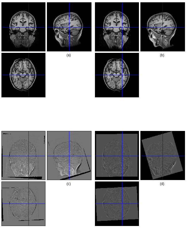
An example of a patient with probable Alzheimer's Disease [anonymised data from the DRC, ION, UCL].
(a) Sections of a 3D T1-weighted MR Image.
(b) The same, from a scan taken one year later, rigidly registered to the first.
(c) A subtraction image indicating structural changes over time.
(d) The determinant of the Jacobian matrix from a high-dimensional registration between the images — brighter colours indicate tissue expansion; darker, contraction or loss.
Analysis of local measures of change (such as the above Jacobian determinants) among a group of subjects, perhaps divided into sub-groups such as patient and control, requires that the different subjects be spatially normalised into correspondence — a challenging task due to the natural anatomical variability in the human brain. Furthermore, statistical analysis, and necessary preprocessing such as data-reduction, should take into account the different factors (or "modes") of variation: spatial, temporal, and individual (perhaps including nested factors such as different subjects within certain sub-groups).
Journal articles
- Ridgway2008b
- Ridgway, G.; Omar, R.; Ourselin, S.; Hill, D.; Warren, J. & Fox, N.
- Issues with threshold masking in voxel-based morphometry of atrophied brains.
- NeuroImage
- 2008
- (Code available)
- Tofts2008
- Tofts, P. S.; Jackson, J. S.; Tozer, D. J.; Cercignani, M.; Keir, G.; Macmanus, D. G.; Ridgway, G. R.; Ridha, B. H.; Schmierer, K.; Siddique, D.; Thornton, J. S.; Wroe, S. J. & Fox, N. C.
- Imaging cadavers: Cold FLAIR and noninvasive brain thermometry using CSF diffusion.
- Magn Reson Med
- 2008, 59, 190-195
- Ridgway2008
- Ridgway, G. R.; Henley, S. M. D.; Rohrer, J. D.; Scahill, R. I.; Warren, J. D. & Fox, N. C.
- Ten simple rules for reporting voxel-based morphometry studies.
- NeuroImage
- 2008, 40, 1429-1435
- Camara2008
- Camara, O.; Schnabel, J. A.; Ridgway, G. R.; Crum, W. R.; Douiri, A.; Scahill, R. I.; Hill, D. L. G. & Fox, N. C.
- Accuracy assessment of global and local atrophy measurement techniques with realistic simulated longitudinal Alzheimer's disease images.
- NeuroImage
- 2008, 42, 696-709
- Henley2008
- Henley, S. M. D.; Wild, E. J.; Hobbs, N. Z.; Warren, J. D.; Frost, C.; Scahill, R. I.; Ridgway, G. R.; MacManus, D. G.; Barker, R. A.; Fox, N. C. & Tabrizi, S. J.
- Defective emotion recognition in early HD is neuropsychologically and anatomically generic.
- Neuropsychologia
- 2008, 46, 2152-2160
- Camara2006
- Camara, O.; Schweiger, M.; Scahill, R.; Crum, W.; Sneller, B.; Schnabel, J.; Ridgway, G.; Cash, D.; Hill, D. & Fox, N.
- Phenomenological Model of Diffuse Global and Regional Atrophy Using Finite-Element Methods.
- Medical Imaging, IEEE Transactions on
- 2006, 25, 1417-1430
Conference papers
- Modat2009
- Modat, M., Ridgway, G., Taylor, Z., Hawkes, D., Fox, N. & Ourselin, S.
- A Parallel-friendly Normalised Mutual Information Gradient for Free-Form Deformation.
- SPIE Medical Imaging
- (Accepted)
- (Please email for pre-print)
- Ridgway2007a
- Ridgway, G.; Camara, O.; Scahill, R.; Crum, W.; Whitcher, B.; Fox, N. & Hill, D.
- Longitudinal Voxel-Based Morphometry with Unified Segmenation: Evaluation on simulated Alzheimer's disease.
- Medical Image Understanding and Analysis
- 2007, 201-205
- (Please email for pre-print)
- Camara2007a
- Camara, O.; Scahill, R.; Crum, W.; Schnabel, J.; Ridgway, G.; Hill, D. & Fox, N.
- Evaluation of local and global atrophy measurement techniques with simulated Alzheimer's disease images.
- Medical Image Understanding and Analysis
- 2007, 16-20
- (MIUA Best Paper Award)
- (Please email for pre-print)
- Camara2007
- Camara, O.; Scahill, R. I.; Schnabel, J. A.; Crum, W. R.; Ridgway, G. R.; Hill, D. L. G. & Fox, N. C.
- Accuracy assessment of global and local atrophy measurement techniques with realistic simulated longitudinal data.
- Med Image Comput Comput Assist Interv
- 2007, 10, 785-792
- Camara-Rey2006
- Camara-Rey, O.; Sneller, B. I.; Ridgway, G. R.; Garde, E.; Fox, N. C. & Hill, D. L. G.
- Simulation of acquisition artefacts in MR scans: effects on automatic measures of brain atrophy.
- Med Image Comput Comput Assist Interv
- 2006, 9, 272-280
Conference abstracts/posters
- Ridgway2008a
- Ridgway, G. R.; Whitcher, B.; Hill, D. L. G. & Fox, N. C.
- Longitudinal Multivariate Tensor- and Searchlight-Based Morphometry Using Permutation Testing.
- 14th annual meeting of the Organization for Human Brain Mapping
- 2008
- (NIH Travel Award)
- Nichols2008
- Nichols, T.; Ridgway, G.; Webster, M. & Smith, S.
- GLM Permutation - Nonparametric inference for arbitrary general linear models.
- 14th annual meeting of the Organization for Human Brain Mapping
- 2008
- Ridgway2007
- Ridgway, G. R.; Scahill, R. I.; Hill, D. L. G. & Fox, N. C.
- A Comparison of Voxel Compression Mapping and Longitudinal Voxel-Based Morphometry.
- 13th annual meeting of the Organization for Human Brain Mapping
- 2007
- Scahill2006icad
- Scahill, R.; Ridgway, G.; Black, R.; Grundman, M.; Hill, D. & Fox, N.
- ICAD IC-104-01 Regional distribution of grey matter changes in Abeta (AN1792) immunized patients with AD: A voxel-based morphometry analysis.
- Alzheimer's & Dementia: The Journal of the Alzheimer's Association
- 2006, 2, 654-654
- Ridgway2006
- Ridgway, G.; Fox, N. & Hill, D.
- Looking for patterns in brain atrophy - principal component analysis of
structural MR images.
- MIAS-IRC Spring-School, Oxford
Other contributions
I frequently offer help on the mailing lists for
SPM
and FSL,
have helped to edit parts of the SPM WikiBook,
and have contributed to
CMIC's internal Wiki.
Ged Ridgway
Centre for Medical Image Computing
Department of Medical Physics
Malet Place Engineering Building
University College London
Gower Street
London WC1E 6BT
United Kingdom
Tel: +44 (0) 20 7679 0218
Fax: +44 (0) 20 7679 0225
Email: Ged(dot)Ridgway(at)gmail(dot)com

